Inquiry Question- How are cells arranged in a multicellular organism?
Unicellular Organisms (Bacteria)
Contain one cell, either prokaryotic or eukaryotic
First forms of life
A single cell carries out all life processes → obtaining nutrients,
exchanging gas, removing waste and reproduction
High SA:V ratio which allows for more efficient movement of
substances - Requires a moist environments for diffusion and osmosis
to occur
Colonial Organisms (Volvox)
A group of cells working or organism working collectively is
called a colony
May be unicellular or multicellular
Can exist independently, however in a multicellular organism
colonial organisms cannot exist alone.
Multicellular Organisms
A community of cells working together to enable the organism to
carry out life processes, including reproduction.
Composed of many different specialised cells, Similar cells are
grouped together and perform specific functions that combine for the
efficient functioning for the organism - Consists of eukaryotic cells.
Large organisms made up of smaller cells increases SA:V ratio.
Each specialised cell type is structurally suited to a particular
function.
Embryonic cells develop suitable structural changes to best suit
their function → Red blood cell
FORMATION OF SPECIALISED CELLS
When cells become specialised they differentiate – they develop
structures enabling them to carry out their function, making
them different to other cells.
Specialised cells originate from stem cells, which are
undifferentiated cells with the ability to divide repeatedly.
[Cell specialisation refers to the function of the cell, while
differentiation is the] [process of a stem cell goes through to
become specialised.]
Enables organisms to grow larger while still efficiently carrying out
processes.
Specialised cells cannot survive independently – they rely on other
cells in the organisms to carry out functions they cannot.
Communication between cells is vital.
In animals this is via the bloodstream and nervous system whereas in
the plants it is brought about by chemical and physical contact
between cells.
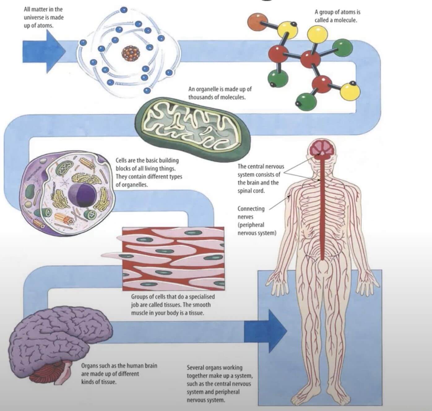
Cell Hierarchy
Organelle Cell Tissue Organ Organ System Organism
Mitochondria Cardiac Muscle Cell Cardiac Muscle Tissue Heart
Cardiovascular System Human
Animal Tissues
Epithelial Tissue
Covers body surfaces, protects organs and forms glands.
Densely packed cells in single sheets or layers.
Doesn’t contain blood vessels.
2 distinct surfaces – exposed to the exterior body cavity or
exposed to adjacent tissue.
Some are specialised for absorption or secretion.
Connective Tissue
Provides support, ensures that all body parts are bound together and
protects against damage
Fibrous connective tissue, loose connective tissue, adipose tissue,
cartilage and bone → Differences are from the arrangement of cells and
specialised structure.
Collagen (strength) + Elastin (Flexibility)
Nervous Tissue
Comprises brain, spinal cord and peripheral nerves.
Highly specialised for communication between all parts of the body
Highly specialised of passing messages between themselves and other
cells
Muscle Tissue
Muscle cells are highly specialized for contraction
Skeletal, Smooth, Cardiac
Responsible for the movement of the body and particular contractions
in various processes (oesophagus peristalsis)
Plant Tissues
Meristematic Tissue
Tips of roots and shoots
Cells divide to produce new growth
Site of cell differentiation
Dermal Tissue
Protects plant tissue and is found in outer layers of stems, roots
and leaves.
Epidermal layer is the outmost, secreting a waxy layer called
cuticle, vital to reduce water loss
Lack Chloroplasts
Vascular Tissue
Responsible for the transport of substances around the plant
Xylem transports water and minerals from the roots to the leaves
Phloem transports products of photosynthesis around the plant
Ground Tissue
Internal cells of a plant other than the vascular
Specialised for storage, support and photosynthesis
Nutrient and Gas Requirements
Inquiry Question: What is the difference in nutrient and gas
requirements between autotrophs and heterotrophs?
Autotrophs
Produce their own organic compounds and energy from inorganic
compounds from their environment, such as carbon dioxide and
water.
Can be divided into two groups:
Photoautotrophs – use light energy (e.g. green plants).
Chemoautotrophs – use chemical energy (e.g. nitrifying bacteria in
the soil)
Heterotrophs
Obtain organic compounds from obtaining other organism
Include all animals and fungi
Vascular and Nonvascular Plants
Majority of autotrophic organisms are plants.
Vascular plants possess a transport system to move substances from
one part of the plant to another.
Plants have specialised cells grouped into tissues
These tissues work collaboratively to carry out life processes like
photosynthesis and gas exchange.
A small number of plants are called non-vascular because they do not
possess this transport system (e.g. mosses and liverworts). - Have a
very simple structure.
All nutrients are absorbed, and wastes are removed by diffusion and
osmosis through the surfaces of the plant.
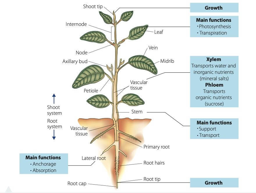
Root System
Usually underground.
The main function of anchoring the plant and absorbing water and
inorganic nutrients from the soil.
Very large surface area.
Absorption occurs through specialised epidermal cells in the
outermost layer of the root.
Increased surface area achieved in the following ways:
Extensive branching (also provides good anchorage)
Root hair zone located in the younger part of each root – epidermal
cells protrude outwards into the surrounding soil, as microscopic
extensions called root hairs.
Flattened epidermal cells increase the exposed surface.
Water moves via osmosis.
Mineral ions usually move via diffusion – if diffusion is too slow,
facilitated diffusion and active transport may be involved.
Root cells have no chloroplasts and thus cannot photosynthesise, but
they can carry out respiration
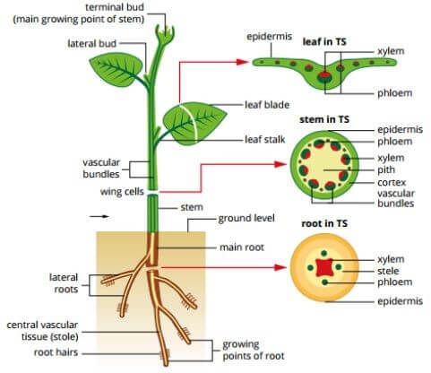
Shoot System (Stem)
Provides structural support and a transport pathway
Located above ground
Consists of 3 main functions
Dermal→ Waterproofing, protection, gas exchange
Vascular → Composed of the xylem and the phloem within vascular
bundles
Ground Tissue → Fills in around vascular tissue
Shoot System (Leaves)
Located above ground
Main function is to absorb sunlight and carbon dioxide and produce
glucose through the process of *photosynthesis.*
Leaves are adapted to absorb the maximum amount of sunlight possible
to provide the energy needed to break bonds in water during the first
stage of photosynthesis.
Thin, flat structure of leaves is well suited to this function – no
internal cell is too far from the light.
Large SA allows maximum absorption.
Transparent epidermis allows sunlight to penetrate the
photosynthetic cells beneath.
Mesophyll is responsible for most of the plant's photosynthesis.
Palisade Cells: Dense with chloroplasts and are main
photosynthetic cells, situated vertically, large numbers ensure
maximum rate of photosynthesis.
Spongy Mesophyll Cells: Irregular in shape and distribution,
situated between palisade cells and lower epidermis, fewer
chloroplasts.
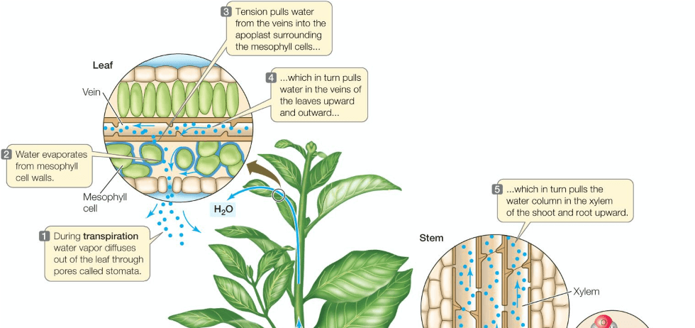
Leaves are also the site of *transpiration,* which is a
process by which water evaporates from the leaf and aids the
movement of water from the roots to the leaves and cools the
plant.
The structure of a leaf allows it to carry out these functions in an
efficient and effective manner.
Sizes and shapes of leaves vary immensely.
Plants in hot, dry habitats have:
Waxy Cuticles – reduce the amount of water lost through
evaporation. - Small Leaves – minimal surface area to reduce water
loss.
Rainforest Plants have:
Large, Thin, Flat Leaves – absorb as much sunlight as possible.
Less concern about water loss due to high humidity.
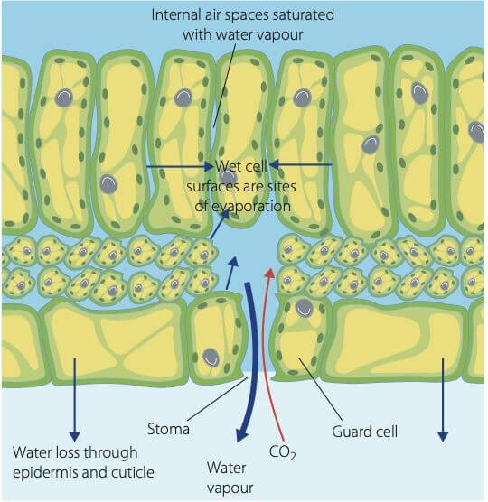
Gaseous Exchange:
Epidermis covers the surface of leaves.
Epidermal cells protect the inner tissues and are able to secrete a
waterproof cuticle to prevent evaporation of water.
Epidermal cells are transparent to allow light to pass to the cell
layers beneath.
Guard Cells – control exchange of gases and the loss of water
through leaves, occur in pairs surrounding the stoma.
Transport
Main transport tissues are the xylem and phloem in the centre of the
root
The main vein in the leaf, the midrib, and many smaller veins branch
out from
it
Distribution of vascular tissue around the plant ensures that all
cells are getting the energy required to function.
Cellular Respiration in Plants
Plants carry out cellular respiration as well as photosynthesis
Occur during night and day
Oxygen and CO2 enter and exit the plant via the guard cells
Nutrient Requirements in Plants
Carbon Dioxide
The opening and closing of the stomata has the greatest effect on
carbon dioxide concentration in the leaf.
If the stomata is closed, available carbon dioxide is used up and
the rate of photosynthesis is reduced.
Water
Amount of water needed for photosynthesis is small compared to that
needed for survival.
When water availability level is low, stomata close and reduce the
amount of carbon dioxide entering the leaf, reducing the rate of
photosynthesis.
Light Energy
The greater the light intensity the faster the rate of
photosynthesis until a plateau is reached.
The plateau is where all photosynthesis systems and enzymes are
working at optimum rate.
Imaging Technologies + Tracing Products Of Photosynthesis
MRI→ uses radio waves and magnetic field to take a series of images
of the plant structures that are used to produce a 3D image of the
structure
X-Ray→ Reveals deeper knowledge of the internal structure of the plant
Radioisotopes are used to determine whether the oxygen released
during photosynthesis originated from the oxygen atom in water or
carbon dioxide
Carbon-14 is added to the carbon dioxide supply of a plant → The
carbon-14 then takes part in the reactions of photosynthesis and is
incorporated into the glucose molecules
The radioisotopes can be traced by the radiation they emit
Gas Exchange in Plants
Leaves are adapted for gas exchange.
Large and flat – large SAV ratio.
Spongy mesophyll layer increases surface area and allow gases to
move freely within the leaf.
Surface of cell is moist
Occurs through stomata and the lenticels.
Stomata:
Found on the underside of the leaf.
Occasionally found on the upper epidermis.
Both sides of the stomata are the guard cells.
These bean-shaped cells contain chloroplasts (unlike other epidermal
cells)
The inner wall of each guard cell is thicker than the outer wall.
Stomata open and close when the guard cells gain or lose water.
Lenticels:
Pores through which gaseous exchange happens in woody plants
Found on trunks and branches of trees and woody shrubs
Appear as small dots, but under the microscope they are seen as
clusters of loose cells in the cork layer
Diffusion through lenticels is relatively slow
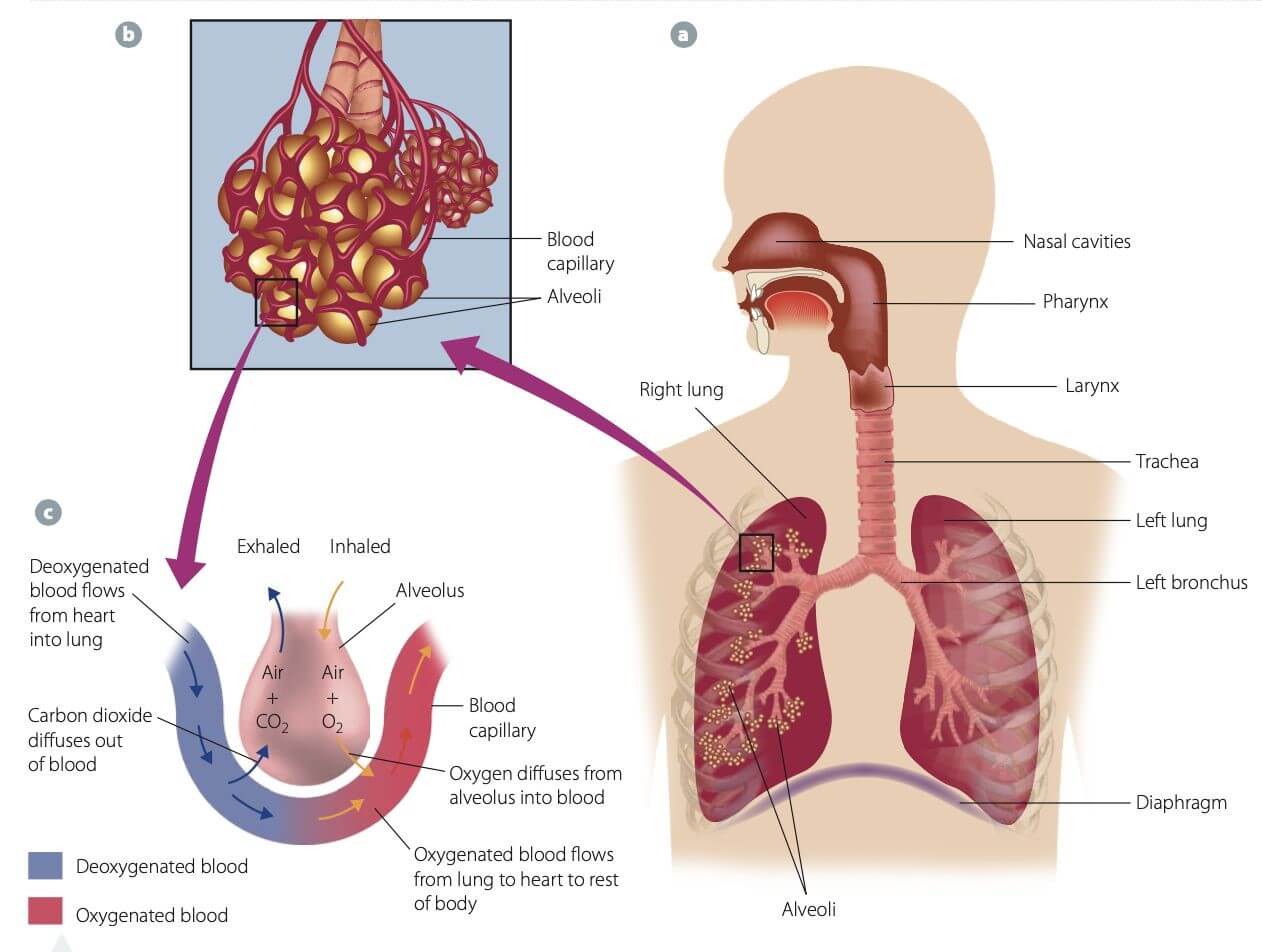
Gas Exchange in Animals
Oxygen is essential for cellular respiration
Carbon Dioxide must be removed as it is highly toxic in large
concentrations - Mammals have lungs, fish have gills and insects have
tracheal system - Large surface area enhanced by folding, branching or
flattening.
Moist, thin surfaces so that gasses can dissolve and diffuse.
Close proximity to the transport system so gases can move easily.
Maintenance of a concentration gradient.
Lungs
Gas exchange structures → alveoli
Increased surface area – folded
Thin lining – flattened single layer of cells
Moist surfaces – saturated with water vapour and mucus
Shares a membrane with the capillaries, hence this facilitates
diffusion of gasses
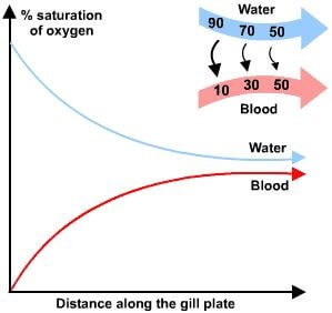
Gills
Gills extract the most oxygen possible out of water
As the water passes through the gills oxygen diffuses into the fish
This is undertaken by a countercurrent process, this ensures the
most oxygen is being diffused from the water (furthest away from
equilibrium).
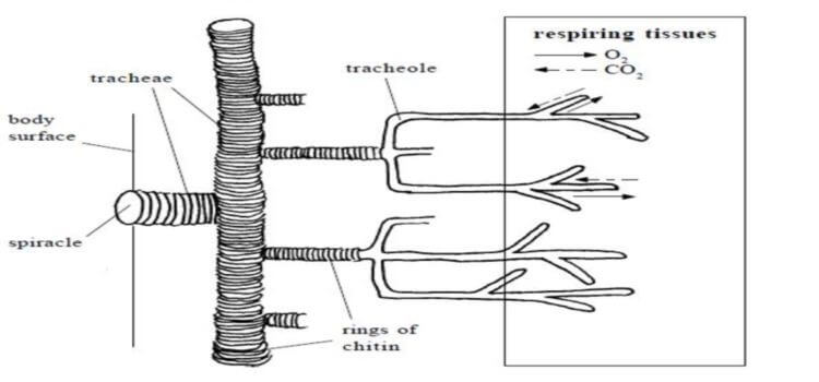
Tracheal System
Insects obtain and release air through spiracles
Do not have lungs or capillaries
Branching air tubes are called tracheal tubes
Oxygen dissolves in fluid, this can be diffused into the cells and
carbon dioxide diffuses out.
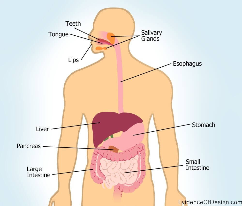
Human Digestive System
Mouth
Teeth break down food for more efficient action of enzymes
Salivary amylase is release and mixed by the tongue
Tongue forms a bolus
Oesophagus
Peristalsis is the muscular contraction that forces food down
Food is moved toward the stomach
Stomach
Gastric juices contain water, HCL and pepsin
Contractions of muscles is a form of mechanical digestion
pH → 2.0-3.0
- Breaks down larger and complex
proteins to a obtainable level
Small Intestine
Emulsifies fats into smaller droplets
Move by diffusion and osmosis
Villi increases the surface area for absorption
Lacteals are collected by the lymphatic system
Glucose and amino acids are absorbed into the capillaries
Liver
Duodenum
Neutralise the acidic chyme leaving the stomach and break down food
Jejunum
Breaks down food into smaller pieces
Breaks down lipids into fatty acids
Ileum
Absorption of products are moved by diffusion or active transport
through villi
Large intestine
Undigested material moves to the large intestine
Site of water and salt absorption
Remaining faeces is moved to the rectum and anus via peristalsis
Transport
Inquiry Question: How does the composition of the transport medium
change as it moves around an organism?
Transport System in Plants
Involves vascular tissue arranged in vascular bundles made up of the
phloem and xylem
Xylem
Moves upwards from the root
Movement upwards from the root.
Consists of xylem tracheids and xylem vessels.
Tracheids: long structures with tapered end walls in contact with
each other.
Xylem vessels are continuous tubes for the transport of water.
- Walls of vessels and tracheids are lined with lignin
– helps prevent the collapse of the vessel and easy movement of
water.
Fibres provide support.
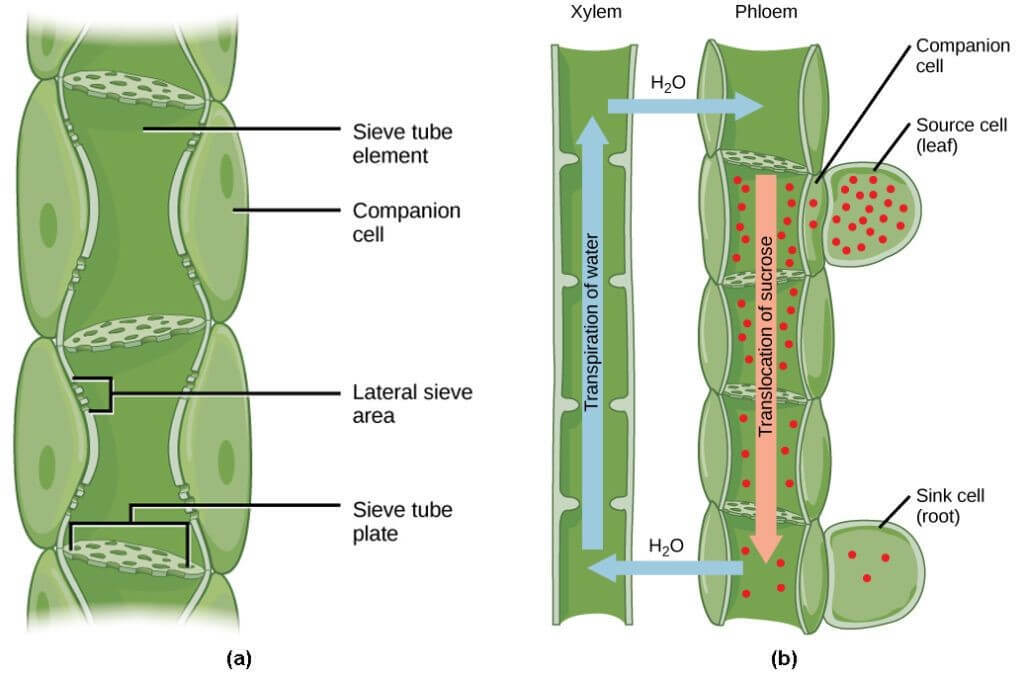
Phloem
Carries products of photosynthesis - Sieve tube cells and companion
cells.
Sieve tube cells are long thin phloem cells with large pores through
their end cell walls.
These perforated cell walls are called sieve plates
Sieve tube cells possess mitochondria and endoplasmic reticulum, but
no nuclei or other organelles
They are arranged end to end forming sieve tubes
Sieve tube cells share cytoplasm, their sieve tubes form channels
through which sugars and other plant products can flow - Companion
cells are found alongside sieve tubes.
They have a nucleus and other organelles that are lacking in sieve
tubes.
Companion cell function is uncertain, but they are thought to assist
effectiveness of sieve tube elements by providing ATP.
They also help with loading and unloading of sugars into a sieve tube.
Transpiration-Cohesion-Tension Theory
Transportation → Process of water vapour leaving the leaves via
the stoma
Cohesion → Water is attracted to itself as it is a polar molecule:
Hydrogen + Oxygen molecules.
Adhesion → Water sticks to the walls of the xylem (Narrow xylem is
more beneficial)
Positive root pressure in the roots via osmosis
Tensions is created by the pull from the leaves
Source-Sink Theory
Glucose produced in the leaf during photosynthesis is either
stored as starch or converted to sucrose and distributed to all
parts of the plant
Distribution is called translocation and occurs in the phloem
Substances in the phloem move in whichever direction is required. -
The phloem also carries amino acids and some mineral nutrients -
Sucrose makes up approx. 90% of phloem sap.
Once it reaches cells is it converted to glucose for respiration or
stored as starch
The movement is driven by the formation of high- and low-pressure
regions within the phloem
Movement occurs from high to low pressure
High-pressure occurs where the sucrose is produced (the source) and
low-pressure occurs where the sucrose is required (the sink)
[The xylem and phloem are adjacent, hence during this process the
water] [from the xylem is diffused into the phloem to dilute the
sugar.] - Actively transported into stem and root cells for
growth.
Transport System in Animals
Open circulation
Found in invertebrates such as insects
Contains one or more hearts that contract to push blood fluid
Hemolymph bathes organs and tissues
Blood and interstitial fluid cannot be distinguished
Blood is in direct contact with tissue
Less efficient, low pressure, Slow
Volume of blood cannot be controlled
Closed Circulation
Found in all vertebrates like fish, mammals frogs and reptiles
Contains blood that is enclosed by blood vessels with a driving
force of the heart
Pathway is from the heart, around the body and back to the heart
Transport nutrients and oxygen to the cells as well as returning
waste and Carbon Dioxide
BLood is not in direct contact with tissue
Heart has 4 chambers to divide oxygenated and deoxygenated blood
Pumped blood can be controlled by contractions and valves
Lymphatic System
Transports excess fluid back into the cardiovascular system and is
made up of lymph vessels and lymph.
Maintains homeostasis
Lymph → Watery fluid
The Heart
Vene Cana → Right Atrium → Right Ventricle → Pulmonary Artery →
Lungs → Pulmonary vein → Left Atrium → Left Ventricle → Aorta
Composed of cardiac muscle cells
Responsible for pumping blood around the body
Pulmonary circulation is blood travelling from heart to lungs
Systemic Circulation is the process of pumping the blood around the
body and back to the heart
Structure of Blood Vessels
Each vessel is best structured to suit the function of the
vessel
Artery
Thicker walls and narrow cross section as blood enters under high
pressure, and thicker walls minimise the chance of the artery tearing.
Walls also are more elastic, so it can expand and contract.
Carries blood from the heart.
Contraction squeezes blood forward and propels it along.
Vein
Thinner walls and wider lumen as the blood is not as high pressure.
The walls are not as elastic as the veins do not need to contract
and expand as much as the arteries.
Returns blood to the heart.
Cross section is wider to allow easy flow of blood.
Blood is propelled by the contracting of muscles surrounding the
veins.
Valves situated at regular intervals to stop the reverse flow of
blood.
Capillaries
Walls are one cell layer thick so that substances can be diffused
efficiently.
Brings blood into close contact with the tissues, enabling exchange
of chemical substances between cells and the bloodstream.
Red blood cells pass through in a single file, increasing their
exposed surface area for the exchange of gases, nutrients and waste.
Blood As a Transport Medium
Red Blood cells (Erythrocytes) - Transport oxygen.
Form in bone marrow.
Haemoglobin (oxygen carrier) is developed within the cell.
Round, biconcave and slightly flattened towards the centre – more
SA:V and elastic in order to squeeze through capillaries.
No nucleus so it has more hemoglobin for oxygen → Structure for
function
White Blood Cells (Leukocytes) - Also produced in bone marrow.
Part of the immune system.
Role is to defend the body against foreign bodies.
Found in tissues as well as the blood.
Can pass through capillaries by squeezing between the cells that
make up the wall of the capillary.
Larger than red blood cells.
Not as abundant as red blood cells.
All white blood cells have a nucleus.
Platelets (Themocytes)
Function in the clotting of blood.
Contact between fibres and platelets causes platelets to break open
and release an enzyme, thromboplastin, which sets in progress a
sequence of steps to seal the blood vessels and cause blood to clot.
Crescent shaped.
Half the size of red blood cells.
Plasma
Yellow, watery fluid.
90% water, 10% protein
Makes up the majority of the volume of blood and carries many
substances throughout the body:
Proteins
Nutrients
Gases
Excretory Waste Products
Ions
Hormones
Vitamins
Changes in Compositions Of Blood
Lungs
As blood moves through the lungs it gains oxygen and loses carbon
dioxide.
Digestive System
Increase in digestive end products.
Lymphatic System
Gain fatty acids that have been emptied into the bloodstream.
Heart
High lipid content.
Stomach
Water and other substances are diffused into the blood.
Liver
Decrease in digestive end products.
Glucose may be added or
removed.
Urea is added to the blood.
Toxins such as alcohol are removed.
Some vitamins and iron are removed.
Kidneys
Urea is decreased.
Excess water and salts are removed.
Large Intestines
Water, salts and vitamins are absorbed into the blood.